


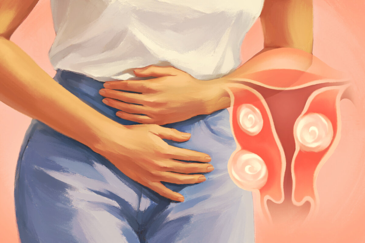
Uterine fibroids are the most common benign tumors of the female reproductive system.
They particularly occur among women in their 30s and 40s, and are estimated to affect up to 70 percent to 80 percent of women by age 50. Yet many never know they have fibroids because they often cause no symptoms, and they are not cancerous.
For others, however, fibroids bring heavy or painful periods and sometimes fertility problems. A range of treatments can relieve symptoms and shrink or remove fibroids.
Symptoms vary depending on the size, location, and number of fibroids (which can grow at different rates), and they usually appear when fibroids interfere with how the uterus works.
Uterine fibroids are classified into three main types based on their location. Subserosal fibroids grow on the outside of the uterus and may press on nearby organs but typically do not affect menstrual flow. Intramural fibroids are the most common type; they develop within the uterine wall and can cause heavy bleeding and pelvic pain or pressure. Submucosal fibroids are less common and grow beneath the uterine lining; they can cause heavy, prolonged bleeding and reduce the chance of pregnancy by blocking the fallopian tubes and interfering with embryo implantation.
Subserosal and submucosal fibroids can be pedunculated, meaning they are attached to the uterus by a thin, stalk-like structure. If a pedunculated fibroid twists on its stalk, it can cut off its blood supply and cause sudden, severe pain.
Common symptoms may include the following:
Period-Related Symptoms
Fibroids can affect menstrual bleeding and cause the following period-related symptoms:
-
Heavy or Prolonged Menstrual Periods: Fibroids developing within the uterine wall (intramural fibroids) may contribute to heavy bleeding by interfering with the uterus’s ability to contract effectively and control blood flow during menstruation. Prolonged periods last eight days or longer.
-
Severe Menstrual Cramps: Fibroids may cause pain by increasing uterine contractions during menstruation or by pressing on nearby organs and nerves.
-
Bleeding Between Periods: Fibroids can cause bleeding outside your usual cycle.
-
Anemia: Ongoing heavy or irregular bleeding can lower your iron and red blood cell levels, leading to fatigue and weakness.
Pressure and Pain Symptoms
Fibroids can press on nearby organs and tissues, causing the following symptoms:
-
A Feeling of Fullness or Pressure in the Lower Abdomen: Larger fibroids can press on nearby organs, such as the bowel, causing constipation or abdominal pain.
-
Frequent Urination: This is caused by larger fibroids pressing on the bladder.
-
Pelvic Pain: Fibroids can cause dull or sharp pelvic pain, which may be more noticeable during menstruation.
-
Pain During Intercourse: Pain may occur only in certain positions or at specific times during the menstrual cycle.
-
Lower Back Pain: Rarely, fibroids can press on the muscles or nerves in the lower back, causing pain.
Fertility Issues
Certain fibroids, especially those that distort the uterine cavity, may reduce the likelihood of conception or successful pregnancy by interfering with fertilization or embryo implantation. In some cases, they can contribute to infertility.
Fibroids may increase the risk of pregnancy complications, including:
-
Placental Abruption: The placenta separates from the uterine wall before delivery.
-
Preterm Labor: Having multiple fibroids or fibroids in contact with the placenta may increase the risk of preterm labor.
-
Repeated Miscarriages: Fibroids, particularly those that distort the uterine cavity, may increase the risk of recurrent miscarriage by interfering with embryo implantation or the developing pregnancy.
-
Delivery Problems: The baby may be in an abnormal position in the uterus, and cesarean birth is more likely.
-
Postpartum Hemorrhage: Excessive bleeding can occur after delivery.
Roughly 15 percent to 30 percent of people with fibroids have severe symptoms. Rarely, fibroids cause emergencies. Get immediate medical care for sudden, severe abdominal pain that does not improve with pain medication, or for heavy vaginal bleeding with lightheadedness, extreme fatigue, or weakness (signs of anemia).
[shortcut_anchor id=”anchor_1792819567716″ label=”Causes”]What Causes Uterine Fibroids?[/shortcut_anchor]The exact cause is not fully understood. Genetic, hormonal, and environmental factors are all thought to play a role, with several theories describing how they work together.
Researchers believe fibroids may begin when a uterine smooth muscle cell, or a stem or progenitor cell, acquires a genetic mutation, most commonly in the MED12 gene. This mutation promotes abnormal cell growth and multiplication.
Studies suggest that estrogen and progesterone stimulate fibroid cell growth and drive production of extracellular matrix, the structural material between cells, which is rich in collagen and fibronectin. As this material builds up, fibroids become firmer and denser. This may help explain why fibroids commonly develop during the reproductive years and often grow during pregnancy, when estrogen and progesterone levels are high.
Growth factors, particularly transforming growth factor beta, may also contribute to fibroid formation and enlargement.
These factors may interact: genetic changes can start the process, while hormones and growth factors help fibroids grow.
Fibroids may grow at different rates during the reproductive years. If they outgrow their blood supply, fibroids can degenerate because of reduced blood flow.
They generally stop growing or shrink after menopause as hormone levels decline. However, they do not always disappear completely. Even when fibroids remain, symptoms such as pelvic pain and heavy menstrual bleeding often improve or resolve after menopause.
Fibroid growth after menopause should be evaluated by a doctor to rule out other conditions, including rare malignancies.
Several factors may increase the risk of developing uterine fibroids, including:
-
Obesity: A 2024 systematic review identified high body mass index as one of the strongest known risk factors for uterine fibroids. Women with obesity may have two to three times the risk of women with average weight. It is unclear whether excess weight directly causes fibroids, but fat tissue can convert androgens into estrogen, potentially promoting fibroid growth.
-
Vitamin D Deficiency: A 2013 study found that women with sufficient vitamin D levels had approximately 32 percent lower risk of developing fibroids than women with insufficient levels. Lab studies using human fibroid cells suggest that active vitamin D may reduce extracellular matrix protein production and inhibit fibroid cell growth.
-
Exposure to Endocrine-Disrupting Chemicals (EDCs): These chemicals can interfere with the body’s hormones. Studies suggest that exposure to certain chemicals, particularly phthalates, may increase fibroid risk and severity, although more research is needed.
-
First Period Before Age 10: Having the first menstrual period before age 10 may increase the risk of uterine fibroids, possibly because it leads to longer lifetime exposure to estrogen and progesterone.
-
No Pregnancy History: Women who have never been pregnant may have an increased risk of developing fibroids due to longer exposure to estrogen and progesterone over multiple menstrual cycles.
-
Age: Fibroids become more common with age, particularly during a woman’s 30s and 40s. Women approaching menopause may have the highest risk of developing fibroids.
-
Race: Uterine fibroids are approximately two to three times more common in black women than in white women. Black women with fibroids also tend to have more and larger fibroids and may experience more severe symptoms. They also tend to develop symptoms about five years earlier on average, although the reasons for these differences are not fully understood.
-
Family History: Having a mother or sister with fibroids raises your risk. A mother with fibroids may increase it about threefold.
[shortcut_anchor id=”anchor_1792819293199″ label=”Diagnosis”]How Are Uterine Fibroids Diagnosed?[/shortcut_anchor]Fibroids are often discovered during a routine pelvic exam or by chance during an ultrasound for another condition. Others are found when women seek care for painful or heavy periods. Only about one-third of fibroids are large enough to be detected during a physical exam, so many may go undiagnosed.
Your doctor will review your symptoms and medical history and perform a physical exam, which may include an internal pelvic exam. Imaging tests can then help confirm the diagnosis.
First-Line Tests
Your doctor may recommend the following tests to diagnose uterine fibroids and assess their effects:
-
Pelvic (Transabdominal) Ultrasound: Uses sound waves, without radiation, to produce images of the uterus and ovaries. The technologist moves a transducer over the abdomen to capture images.
-
Transvaginal Ultrasound: Involves gently inserting a special ultrasound probe into the vagina after you empty your bladder. The procedure is usually not painful and provides detailed images of the uterus, uterine lining, and ovaries.
-
Magnetic Resonance Imaging (MRI): Provides more detailed images of the number, size, and location of uterine fibroids than ultrasound. MRI may be used when ultrasound findings are unclear or when detailed mapping is needed before treatment. Depending on the clinical question, MRI may be performed with or without intravenous contrast.
-
Blood Tests: Identify fibroid complications rather than diagnose the growths themselves. A complete blood count checks for anemia by measuring red blood cells and hemoglobin, while iron tests show whether iron deficiency is contributing.
Specialist Tests
Your doctor may refer you to a gynecologist for additional tests, particularly if imaging findings are unclear or you have abnormal bleeding or fertility concerns. These tests may include:
-
Saline Infusion Sonogram (Saline Hysterosonography): A small catheter is passed through the cervix to fill the uterus with sterile saline, allowing ultrasound to capture detailed images of the uterine cavity and lining to identify abnormalities such as submucosal fibroids and polyps.
-
Hysteroscopy: A thin telescope is inserted through the cervix, and sterile saline or carbon dioxide gas is used to expand the uterine cavity for a better view. This procedure can detect submucosal fibroids and uterine polyps.
-
Laparoscopy (Keyhole Surgery): A laparoscope (thin telescope) is inserted through a small incision in the abdomen to examine the reproductive organs. It is not routinely needed to diagnose fibroids.
-
Endometrial Biopsy: Involves taking a small sample of the uterine lining to check for cancer or other abnormalities, particularly in cases of abnormal uterine bleeding.
-
Hysterosalpingography: A specialized X-ray procedure used to detect abnormalities in the size and shape of the uterus and fallopian tubes.
Once diagnosed, most uterine fibroids do not require treatment unless they cause symptoms or complications.
[shortcut_anchor id=”anchor_1792819229942″ label=”Treatments”]What Are the Treatments for Uterine Fibroids?[/shortcut_anchor]Because more than 99 percent of fibroids are noncancerous and do not increase the risk of uterine cancer, for women without symptoms, actively monitoring fibroids without treatment may be an appropriate option.
For fibroids that do need treatment, options depend on their size, number, location, and the patient’s preferences, including whether a woman wants to become pregnant in the future. Treatment aims to reduce menstrual bleeding, relieve pain and pressure, improve bladder and digestive function, and preserve or improve fertility.
Medications
Medications can temporarily relieve fibroid symptoms but do not eliminate fibroids. They may help manage heavy bleeding before surgery, but they are generally ineffective for pressure symptoms caused by large fibroids.
-
Nonsteroidal Anti-Inflammatory Drugs (NSAIDs): Medications such as ibuprofen or naproxen may reduce menstrual bleeding and relieve fibroid-related pain, making them a conservative option for occasional pelvic discomfort.
-
Tranexamic Acid: Can reduce heavy menstrual bleeding by up to 40 percent. It also prevents blood clots from breaking down too quickly.
-
GnRH Agonists: Stimulate and then suppress the release of luteinizing hormone (LH) and follicle-stimulating hormone (FSH), reducing ovarian estrogen production. The temporarily lowered estrogen levels can shrink fibroids, stop periods, and improve blood counts before surgery. Because of side effects such as hot flashes and bone loss, they are generally used short-term.
-
Oral GnRH Antagonist Combination Therapies: Elagolix and relugolix are available in combination with low-dose estrogen and progestin. These medications reduce heavy bleeding, while the added hormones help limit hot flashes and bone loss. Treatment is generally limited to 24 months because continued bone loss may be irreversible.
-
Hormonal Contraceptives: Combined birth control pills, the patch, and the vaginal ring contain estrogen and progestin and may reduce fibroid-related bleeding. Women aged 35 and older who smoke are generally advised to avoid estrogen-containing contraceptives because of the higher risk of blood clots and cardiovascular problems.
-
Selective Estrogen Receptor Modulators: May help slow fibroid growth, but their effectiveness in relieving symptoms remains unclear.
-
Iron Supplements: Used to prevent or manage anemia caused by heavy menstrual bleeding.
Progestin-Releasing IUD (Intrauterine Device)
This small device releases progestin into the uterus, thinning the uterine lining and reducing heavy and painful menstrual bleeding. It is suitable for women whose fibroids do not distort the uterine cavity, but it does not shrink or eliminate the fibroids themselves.
Procedural Treatments
These minimally invasive interventions reduce fibroid size, block fibroids’ blood supply, or relieve symptoms while potentially preserving the uterus.
-
Uterine Artery Embolization: Using X-ray guidance, a catheter is inserted through an artery in the groin to deliver tiny embolic particles, such as polyvinyl alcohol particles or tiny gelatin-based microspheres, that block the blood supply to the fibroids, causing them to shrink and die. It can reduce heavy menstrual bleeding, pelvic pain and pressure, frequent urination, and constipation.
-
Magnetic Resonance-Guided Focused Ultrasound: Uses focused sound waves, guided by MRI, to heat and shrink fibroids, particularly those located at the front of the uterus. Its effects on fertility are not yet fully understood.
-
Radiofrequency Ablation: Uses laparoscopy and ultrasound guidance to deliver heat energy that shrinks and softens fibroids. Its effects on fertility are not yet fully understood.
-
Endometrial Ablation: Destroys the lining of the uterus to reduce or stop heavy menstrual bleeding, but menstruation often stops completely. It may help women with small fibroids but does not remove the fibroids themselves. It is not suitable for women who want future pregnancies, and contraception is still needed because pregnancy after ablation can be dangerous.
-
Myolysis: Destroys fibroid tissue using an energy source such as a laser, extreme cold (cryomyolysis), a heated electrical needle, or high-intensity focused ultrasound energy. It is generally used for smaller fibroids and is not recommended for women planning future pregnancies because of the risk of uterine scarring and infection.
Surgeries
Surgery either removes fibroids while keeping the uterus—myomectomy—or removes the uterus entirely—hysterectomy. In a 2024 clinical review published in the New England Journal of Medicine, researchers called for using less-invasive treatments when possible for better health outcomes. Hysterectomy carries risks and potential long-term consequences, and the benefits and risks vary from person to person.
Myomectomy: Removes uterine fibroids while preserving the uterus, making it a suitable option for women who wish to maintain their fertility. After myomectomy, the risk of recurrence is higher in younger women and those who have more fibroids at the time of surgery. However, a previous myomectomy may lead to a recommendation for cesarean delivery, because removing fibroids can weaken the uterine wall.
The three main types of myomectomy are:
-
Abdominal Myomectomy: Fibroids are removed through an incision in the lower abdomen, often called a “bikini cut,” and the uterine muscle is stitched back together. This type has the longest recovery period, typically four to six weeks.
-
Laparoscopic Myomectomy: Fibroids are removed through several small abdominal incisions using a camera and specialized instruments, with a shorter recovery period of two to four weeks.
-
Hysteroscopic Myomectomy: Submucosal fibroids are removed through the vagina and cervix using a thin telescope and specialized instruments, without abdominal incisions. This type has the shortest recovery period, typically one to four days.
Hysterectomy: Fibroids are the most common reason for hysterectomy in the United States. This procedure removes the uterus, permanently resolving fibroid-related menstrual bleeding and preventing fibroids from recurring. The ovaries may also be removed to reduce the risk of ovarian cancer or to treat certain ovarian conditions, although removal is not routinely necessary for fibroid treatment. The decision to remove the ovaries or not depends on menopausal status, medical conditions, and personal preferences, because removing the ovaries before menopause can cause symptoms such as hot flashes and vaginal dryness. A total hysterectomy removes both the uterus and cervix, while a subtotal hysterectomy removes the uterus but leaves the cervix in place.
The three main approaches to hysterectomy are:
-
Vaginal Hysterectomy: The uterus is removed through the vagina without an abdominal incision. This approach is suitable when the uterus is not too large.
-
Abdominal Hysterectomy: The uterus is removed through an incision in the lower abdomen.
-
Laparoscopic Hysterectomy: The uterus is removed through small abdominal incisions using a camera and specialized instruments. This approach may not be suitable for women with very large fibroids or an enlarged uterus.
Herbs and Herbal Preparations
Current evidence is insufficient to support or refute the use of herbal preparations for treating uterine fibroids, owing to a lack of large-scale, high-quality clinical studies. However, preliminary findings from pilot studies and small clinical trials suggest potential benefits. Consult your healthcare provider before using any of the herbs or preparations below.
Cinnamon and Poria Formula (Guizhi Fuling): A 2023 meta-analysis of 28 studies involving more than 2,800 patients found that cinnamon and poria formula combined with low-dose mifepristone, a medicine that blocks progesterone, significantly reduced fibroid and uterine volume, menstrual flow, and reproductive hormone levels compared with mifepristone alone. Another 2014 systematic review of 38 studies involving nearly 4,000 participants found that cinnamon and poria formula, particularly when combined with mifepristone, may reduce uterine fibroid volume and alleviate period cramps.
Bur-Reed Rhizome: A 2025 meta-analysis of 19 studies found that Chinese herbal medicines combined with high-intensity focused ultrasound (HIFU) significantly reduced uterine fibroid volume and improved clinical response rates compared with HIFU alone, with bur-reed rhizome (Sparganii rhizoma) among the most frequently used herbs.
Acupuncture
Acupuncture may help relieve fibroid-related pain and other symptoms, but more high-quality clinical trials are needed to confirm its effectiveness and safety in treating uterine fibroids.
A 2023 meta-analysis of 11 studies found that acupuncture may reduce fibroid and uterine volume, improve symptoms, and increase treatment effectiveness, with no serious adverse effects reported.
Acupuncture With Chinese Herbs: A 2024 meta-analysis of nine studies involving 640 women with uterine fibroids found that combining Chinese herbal medicines with acupuncture improved overall treatment response rates and reduced fibroid size compared with Chinese herbs alone.
Tung’s Acupuncture: A 2019 feasibility study evaluated a seven-point Tung’s acupuncture protocol for uterine fibroids in nine participants who received acupuncture twice weekly for six or 12 weeks. Eight participants were included in the final symptom analysis, and all reported improvements. After 12 weeks, nine symptoms, including heavy bleeding, lower back pain, and nausea, had resolved completely.
[shortcut_anchor id=”anchor_1792819006611″ label=”Lifestyle Approaches”]What Are the Natural and Lifestyle Approaches to Uterine Fibroids?[/shortcut_anchor]Self-care and lifestyle changes can ease symptoms and support overall health.
1. Self-Care Strategies
The following strategies may help relieve fibroid-related pain and discomfort.
-
Heat: Applying a heating pad or hot water bottle to your lower abdomen or taking a warm bath may relax muscles and ease pain.
-
Comfortable Positions: Lying on your back with a pillow under your knees or on your side with your knees drawn toward your chest may ease pain and reduce pressure on the back and pelvis.
-
Regular Exercise: Engaging in aerobic exercise may improve circulation and stimulate the release of endorphins, the body’s natural pain relievers.
2. Supplements
It’s best to check with your healthcare provider before trying any of the following supplements, especially if you take medications or have an underlying health condition.
-
Curcumin: This compound in turmeric has anti-inflammatory and antioxidant properties. A small uncontrolled 2013 study of 50 women reported improvements in bleeding and fibroid measurements after curcumin treatment. However, the study lacked a control group, and its findings have not been confirmed in rigorous clinical trials. Another uncontrolled study found that average fibroid diameter decreased slightly, from 4.74 to 4.46 centimeters. Without a comparison group, neither study can establish that curcumin caused these changes.
-
Vitamin D: A 2024 meta-analysis of five studies involving 511 participants found that vitamin D supplementation reduced uterine fibroid size compared with a placebo, especially when taken for more than eight weeks. However, larger, well-designed clinical trials are needed to confirm its effectiveness. A 2019 study of 69 women with uterine fibroids and vitamin D deficiency found that those receiving 50,000 international units of vitamin D every two weeks had increased blood vitamin D levels and reduced fibroid size compared with the placebo group.
-
Green Tea Extract: In a 2013 study of 39 women with symptomatic fibroids, those who took green tea extract for four months saw their total fibroid volume fall by 32.6 percent, while the placebo group’s rose by 24.3 percent. Bleeding and other symptoms also improved. Concentrated green tea extracts have been linked to rare cases of liver injury, so use caution, particularly if you have liver disease.
-
Combined Supplements: A 2021 study of 95 women of childbearing age with fibroids smaller than 4 centimeters found that the 41 who took vitamin D, epigallocatechin gallate (EGCG), and vitamin B6 for four months had a 38 percent reduction in total fibroid volume, while volume rose in the untreated group. A 2025 study of 33 women taking the same combination for 16 weeks found that 94 percent experienced a 12.7 percent average reduction in fibroid volume. Symptoms and quality of life improved, although bleeding did not significantly improve.
3. Diet
A 2024 narrative review suggested emphasizing fruits, vegetables, whole grains, and other minimally processed foods while limiting highly processed foods to support overall health and possibly reduce fibroid risk or progression. However, no specific diet has been proven to slow the growth of existing fibroids.
4. Yoga
A 2024 study of 42 women aged 25 to 45 years with uterine fibroid symptoms found that after practicing yoga for one hour daily, five days a week, for 90 days, the participants experienced reductions of 38 percent in uterine fibroid measurements, 34 percent in endometrial thickness, 47 percent in right ovarian measurements, and 32 percent in left ovarian measurements.
A three-month 2025 experimental study of 20 perimenopausal women aged 40 to 50 with uterine fibroids examined the effects of physio-yoga, combining aerobic exercise and resistance training. The intervention improved pelvic pain, body mass index, and menstrual regularity.
[shortcut_anchor id=”anchor_1792818524895″ label=”Prevention”]How Can I Prevent Uterine Fibroids?[/shortcut_anchor]There is no proven way to prevent uterine fibroids. However, the following factors may help reduce the risk of developing them, though most of the evidence comes from observational studies, which show associations rather than cause and effect.
-
Diet: A balanced diet rich in fruits and green vegetables, with limited red meat and processed foods, may be associated with a lower risk of developing fibroids. A 2024 systematic review of 33 human studies found that high fruit and vegetable intake and adequate vitamin D intake were the strongest potential protective factors against fibroids.
-
Adequate Vitamin D: A 2013 study of more than 1,000 premenopausal women aged 35 to 49 found that women with sufficient vitamin D levels had a 32 percent lower risk of developing uterine fibroids than women with insufficient levels. Greater sun exposure was also associated with a lower likelihood of having fibroids. A pilot trial found that vitamin D supplementation did not significantly reduce fibroid recurrence after surgery, although recurrent fibroids were smaller among participants receiving vitamin D. Larger trials are needed to confirm these findings.
-
Higher-Intensity Exercise: Regular physical activity helps maintain a healthy weight and may contribute to a lower risk of fibroids. A 2025 systematic review of 15 studies found no significant link between regular physical activity and a lower risk of uterine fibroids. However, higher-intensity physical activity was associated with a slightly lower likelihood of having fibroids.
-
Herbal Products: A 2019 study of 52,151 Taiwanese women aged 20 to 45 found that Chinese herbal product use was associated with a lower incidence of uterine fibroids compared with nonuse, with an even lower incidence among users of phytoestrogen-containing products.
-
Reduced Chemical Exposure: A 2024 meta-analysis of 14 observational studies involving nearly 6,000 women found that higher exposure to phthalates, chemicals that make plastics flexible and durable, was associated with higher odds of uterine fibroids. The authors of a 2024 review suggested limiting endocrine-disrupting chemicals by avoiding heating food in plastic containers, limiting single-use plastics, choosing fragrance-free personal care products, dusting regularly, washing hands often, minimizing pesticide exposure, and choosing fresh, minimally processed foods over heavily packaged ones.
-
Childbirth: Having given birth to multiple children has been associated with a lower risk of fibroids, although this is not considered a preventive intervention. This may be due to changes in reproductive hormone exposure and uterine tissue remodeling after childbirth, although the exact mechanisms remain unclear.
[shortcut_anchor id=”anchor_1792818359880″ label=”Complications”]What Are the Possible Complications of Uterine Fibroids?[/shortcut_anchor]Most fibroids do not cause serious complications, but some cause problems that go beyond everyday symptoms. Possible complications include:
-
Anemia: Heavy menstrual bleeding can cause anemia, the most common complication of uterine fibroids.
-
Severe Pain or Heavy Bleeding: These symptoms may require emergency medical treatment or surgery in severe cases.
-
Abdominal Swelling: Very large fibroids may cause abdominal swelling.
-
Urinary Tract Infections: Fibroids that press on the bladder may prevent it from emptying completely, increasing the risk of infection.
-
Hydronephrosis: A fibroid growing toward the side of the uterus can compress the ureter, the tube that carries urine from the kidney to the bladder. This can block urine flow and cause the kidney to swell.
-
Fibroid Torsion: A fibroid attached to the uterus by a stalk can twist, cutting off its blood supply and causing sudden, severe pain that may require emergency surgery.